Bochdalek hernia radRounds Radiology Network
We diagnosed incidental Bochdalek's hernias in 22 patients on the basis of radiology case reviews (Figs. 1,2,3). In each instance, the patient's symptoms were not directly referable to the site of, or contents within, the hernia, and so we deemed the finding of the hernia to be incidental.

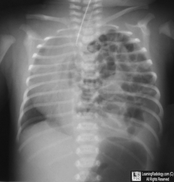
Neonatal Bochdalek hernia. a A 1dayold boy with left Bochdalek... Download Scientific Diagram
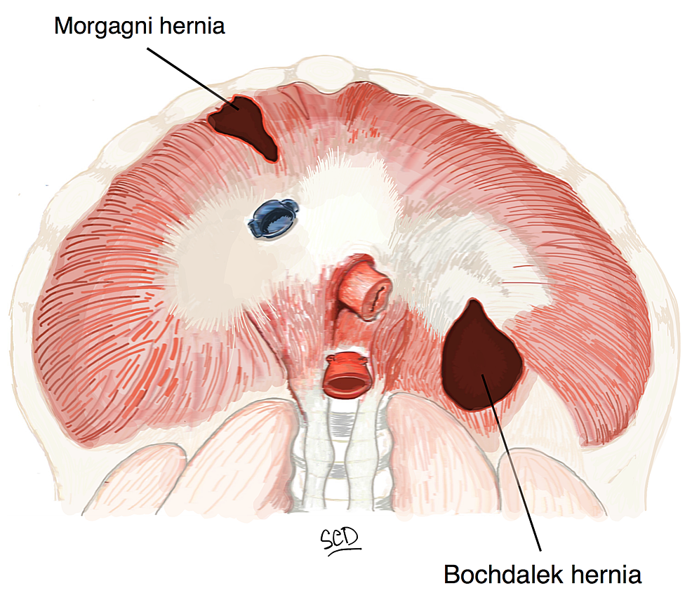
Bochdalek hernia is one of two forms of a congenital diaphragmatic hernia, the other form being Morgagni hernia. A Bochdalek hernia is a congenital abnormality in which an opening exists in the infant's diaphragm, allowing normally intra-abdominal organs (particularly the stomach and intestines) to enter into the thoracic cavity..

Bochdalek hernia Radiology Case Radiologic Technology, Radiology
Bochdalek hernias are often incidental findings on imaging studies or discovered in the workup of respiratory distress, whereas hiatal hernias are typically diagnosed through endoscopic evaluation. An adult Bochdalek hernia is rare and can present as a result of the iatrogenic weakness of the diaphragm due to major surgery.

Rightsided Bochdalek’s hernia in an adult The American Journal of Surgery
The chest and abdominal computed tomography (CT) scans of 940 patients were reviewed to determine the prevalence of Bochdalek hernias and to evaluate the widely held concept that left-sided hernias occur more than nine times as often as right-sided hernias. Sixty Bochdalek hernias were identified in 52 patients, a prevalence of 6%, which is more than 100 times more frequent than previously.
Cureus Diaphragmatic Hernia Repair Using Biosynthetic Tissue Reinforcement Patch A Case
Posterior - Bochdalek. Most common. Occurs through old pleuroperitoneal canals. Just lateral to the spine on either side. More frequent on left side. Possibly due to "protection" of right-side by liver. Hernia may contain intestine, stomach, spleen, liver or omentum. If hernia occurs on right. Intestine and liver or only liver may herniate.
Bochdalek hernia radRounds Radiology Network
Bochdalek Hernia. More common. Occurs through old pleuroperitoneal canals. Just lateral to the spine on either side. More frequent on left side. Possibly due to "protection" of right-side by liver. Hernia may contain intestine, stomach, spleen, liver or omentum. If hernia occurs on right. Intestine and liver or only liver may herniate.

Pin on Paediatric Radiology
Journal of Thoracic Imaging, Vol. 24, No. 1 Prevalence of Incidental Bochdalek's Hernia in a Large Adult Population November 23, 2012 | American Journal of Roentgenology, Vol. 177, No. 2
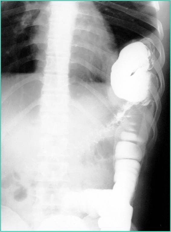
Scaphoid Abdomen Diaphragmatic Hernia A small diaphragmatic hernia is diagnosed by fluoroscopy
Bochdalek hernia is a developmental defect in the posterolateral diaphragm, allowing herniation of abdominal contents into the thorax, causing mechanical compression of the developing lung parenchyma and sometimes causing lung hypoplasia. As such, symptoms typically manifest in the pediatric age group and tend to be respiratory. Symptomatic adults are diagnosed rarely, with the majority of.

De 25+ bedste idéer inden for Pediatric radiology på Pinterest Scrubs ensartet, Nclex og
Citation, DOI, disclosures and article data. Bochdalek hernias , also known as pleuroperitoneal hernias, (alternative plural: herniae) are the commonest type of congenital diaphragmatic hernia. They occur posteriorly and are due to a defect in the posterior attachment of the diaphragm when there is a failure of pleuroperitoneal membrane closure.
Bochdalek (Pleuroperitoneal) Hernia radRounds Radiology Network
Imaging description. A Bochdalek hernia is a defect of the posterior hemidiaphragm with protrusion of abdominal content, usually fat, into the thorax [1]. It may occur on either side, but is more common on the left side due to a protective barrier effect of the liver [1, 2]. CT typically demonstrates the diaphragmatic defect with abdominal fat.

17 Best images about Radio Imaging Chest on Pinterest Pulmonary edema, Signs and Popcorn
Patient Data. Age: 20 years. Gender: Female. ct. Defect in posteromedial aspects of right hemi-diaphragm with herniation of large, small bowel and right kidney into thorax.

Bochdalek hernia The Lancet
Coronal C+ portal. venous phase. Sagittal C+ portal. venous phase. CT scout image reveals indistinct right diaphragmatic copula with right paracardiac soft tissue shadow. CT images show a defect of the right crus of the diaphragm with herniation of the stomach, the first part of the duodenum and part of the left lobe of the liver into the right.

Congenital diaphragmatic hernia Image
Search 33 Cingoli landscape architects & designers to find the best landscape architect or designer for your project. See the top reviewed local landscape architects & designers in Cingoli, The Marches, Italy on Houzz.

Bochdalek hernia Radiology Case
Characteristics. Congenital anomaly with defective fusion of the posterolateral pleuroperitoneal layers. 85-90% on the left, 10-15% on the right. Usually unilateral lying posteriorly within the chest. Hernia may contain fat or intra-abdominal organs. In neonates the hernia may be large and present in utero.

Bochdalek Hernia images, diagnosis, treatment options, answer review Thoracic Imaging
Bochdalek hernia is usually left sided and maybe an incidental finding in as high as 10% of asymptomatic adult 1 . As demonstrated in this case, the herniation content is usually retroperitoneal fat and less often kidney (right sided). Anteroposterior chest radiograph shows double density of right hemidiaphragm.

CT scan showing anterior left diaphragmatic hernia with small and large... Download Scientific
The patient was diagnosed with Bochdalek hernia based on the presence of a hernia orifice in the posterolateral aspect of the left diaphragm. When CT imaging performed 10 years before was retrospectively reviewed, a more modest degree of Bochdalek hernia was detected, which may have caused the gastric ulcer at that time (Fig. 2 ).
